Picturing Knowledge: Scientific Images in Printed Books (1450–1800)

Exploring the Human Body

Exploring the Human Body

Maya Caselnova, Ben Miller, and Kaeo Wongbusarakum

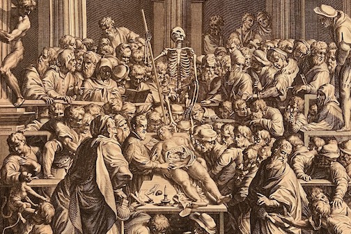
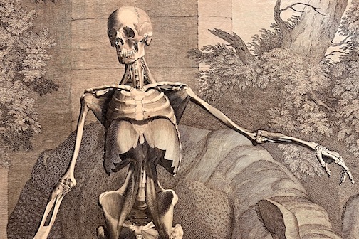
In order to understand the world around us, we must first understand ourselves. The discipline of anatomy represents the response to this innate human desire to understand and offers a glimpse into the fabric of our own biology. This broad field seeks to provide insight into the functions and organization of the human body. In the early stages of anatomical exploration, as with any scientific study, there was great discord surrounding the proper ways to conduct dissections and disseminate knowledge. Preliminary studies of human anatomy and surgery involved “the professor [reading] out the text of Galen while a barber-surgeon cut open the body.”[1] This style of dissection highlights early anatomical exploration as heavily reliant upon canonical texts and the use of intellect, rather than experience and understanding. Andreas Vesalius (1514-1564) revolutionized this field when “he advocated firsthand dissections” and “ocular faith of witnesses in the dissection hall through pictures.”[2] These efforts aided the development of anatomy into a more objective and tangible area of study that led to an intimate understanding of ourselves and the fabric from which we are constructed. These books, while more scientifically focused than prior studies of anatomy, still included extravagant portraits of humanized skeletons and animated corpses — embellishments that are rarely found in the pages of modern anatomy textbooks. Vesalius’ insistence on including images in books and advocating that physicians personally complete dissections was met with great resistance, “and [the] use of pictures did not become the norm in anatomical works even in the eighteenth century.”[3] Vesalius’ work has been described by scholars as a “pictorial form of argument.”[4] Through this exhibit, you will gain an understanding and appreciation for the complex arguments made through pictures in not only Vesalius’ work but the work of a curated group of influential physicians.
Case 8: Through Flesh and Bone

During the 1500s there were contentious debates among physicians about the proper methods for exploring and understanding human bodies. A new method based on dissections and experiential studies renewed the study of anatomy, providing glimpses into the normally unseen and intangible insides of the human body. For the first time, people were able to visualize and appreciate these hidden interior features. This case explores three examples showing the expansion of anatomical understanding and the methods by which these discoveries were made. (Maya Caselnova, Ben Miller, and Kaeo Wongbusarakum)

Click to see an item.
Engraved frontispiece to Andreas Vesalius, Opera omnia anatomica & chirurgica (Leiden, 1725)
Side view of the human skeleton in Andreas Vesalius, Opera omnia anatomica & chirurgica (Leiden, 1725)
“Organi Visus,” in Giulio Cesare Casseri, Pentaestheseion (Venice, 1609)
During the Renaissance, Andreas Vesalius advanced the art of anatomical illustrations from simple, five-figure series to more accurate, naturalistic representations of the human body. Influenced by Greek and Roman statues and the newly discovered laws of perspective, he depicted aesthetically pleasing figures posed dramatically with classical architecture in the background.[5] This was a major change from the first medical illustrations, which used bare backgrounds and bland poses. Part of this shift could be attributed to the increase in nobility and social status surrounding the study of anatomy in northern Italy.[6] As a humanist, Andreas Vesalius recommended that his students read anatomy texts by the ancient Greek physician Galen; however, he asserted that learning about anatomy by reading books was insufficient. Through meticulous dissections, Vesalius corrected the teachings of Galen, who was unable to dissect cadavers due to his religious beliefs.[7] Vesalius believed demonstrable evidence took precedence over written text, and thus unified dissection with lecturing, asserting that hands-on investigation was the best way to acquire knowledge about the human body. Vesalius’ landmark publication, De humani corporis fabrica (Basel, 1543), based the acquisition of anatomical knowledge on dissection. It communicated this knowledge about the human body through illustrations, as opposed to written descriptions, reinforcing the tactile and visual components of learning about human anatomy. Furthermore, because Vesalius taught at Padua University in northern Italy during the Renaissance, he had access to some of the most talented artists. Fabrica features an array of woodcut illustrations illustrating complex attitudes about human anatomy. Vesalius most likely chose Jan van Calcar to illustrate Fabrica, because he was a painter from the Titian school, which focused on the vivid representation of nature and human emotions.[8] By mixing approaches from art and science, the book’s anatomical illustrations are both gripping and disturbing. They often humanize cadavers by including features like facial hair alongside half-peeled, gaping holes revealing the deceased models’ brains.
Case 9: The Art of Anatomy

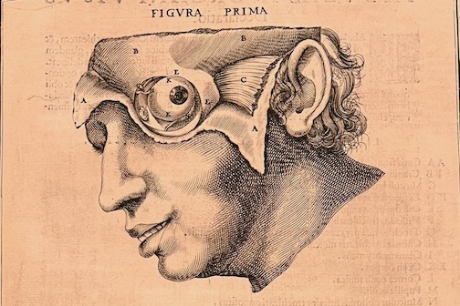
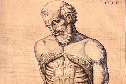
Since most people were not able to attend dissections, images played an instrumental role in allowing readers of anatomy books to interact with their contents and discoveries about the human form. This reliance on images meant that early modern authors took liberties in artistic representation and depicted canonical bodies in keeping with the idea of a perfect human form. Their pursuit of perfection was synonymous with portraying God’s creation in its ideal form. This case presents two works that present anatomical knowledge through stylized artistic images. (Maya Caselnova, Ben Miller, and Kaeo Wongbusarakum)

Click to see an item.
“Fourth Muscular Table,” in Bernhard Siegfried Albinus, Tables of the Skeleton and Muscles of the Human Body Translated from the Latin (London, 1749)
Abdominal muscles, in Adriaan van de Spiegel, De humani corporis fabrica libri decem (Venice, 1627)
Anatomy was not only the study of the human body but also the study of God's creation. While during the Middle Ages the study of the human body was not given great importance, the Renaissance brought about a newfound interest in studying the inner workings of the human body. Vesalius described the human body as being a fabrica, a Latin term that refers to an object that is fabricated or fashioned by an intelligent creator.[9] Within Vesalius’ Fabrica, he is depicted in a portrait studying a dissected hand. He describes how the intricate interlacing of tendons and muscles could only be present through the influence of an “almighty creator” with “outstanding ingenuity.”[10] If we also look at Renaissance art, we see that painters and sculptors placed heavy emphasis on the human body as a creation of God. In Rembrandt’s “The Anatomy Lesson of Dr. Nicolaes Tulp,” a painting depicting the dissection of a hand, we can once again see the argument that complexity implies creative and intelligent design.[11] By focusing on the hand, the artwork draws attention to the hand and body as God's creation and that it is also through this creation that humans are given the power to study the human body.[12] It is the complexity of the body created through God’s wisdom that Renaissance physicians and artists strove to understand and express through their works. Through the act of studying God's creation, anatomists like Vesalius as well as artists created an idea of the ideal body or the canonical body.[13] This “ideal body” did not necessarily represent human anatomy as it actually was, but rather as it was desired and “created” to be. The study of anatomy at this time was in part driven by a desire to understand God’s creation in all of its complexity and peculiarity.


[1] Sachiko Kusukawa, Picturing the Book of Nature: Image, Text, and Argument in Sixteenth-Century Human Anatomy and Medical Botany (Chicago: The University of Chicago Press, 2012), 202.

[2] Kusukawa, Picturing the Book of Nature, 248.

[3] Kusukawa, Picturing the Book of Nature, 233.

[4] Kusukawa, Picturing the Book of Nature, 227.

[5] Alan Branigan, “History of Medical Illustration” (Association of Medical Illustrators, Illuminating the Science of Life, 1995).

[6] Kris Newby, “Stanford art students get lesson on the evolution of anatomy illustration,” (Stanford Medicine, 2017).

[7] Fabio Zampieri et al., “Andreas Vesalius: Celebrating 500 years of dissecting nature” (Global Cardiology Science and Practice, 2015).

[8] Fabio Zampieri et al., “Andreas Vesalius: Celebrating 500 years of dissecting nature”.

[9] Kusukawa, Picturing the Book of Nature, 215.

[10] Kusukawa, Picturing the Book of Nature, 204.

[11] Dolores Mitchell, “Rembrandt’s ‘The Anatomy Lesson of Dr. Tulp’: A Sinner among the Righteous,” Artibus et Historiae 15, no. 30 (1994): 147.

[12] Mitchell, “Rembrandt’s ‘The Anatomy Lesson of Dr. Tulp,” 147.

[13] Kusukawa, Picturing the Book of Nature, 221.

Contents of this path: