X-Rays
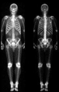
The process of medical science is dependent on somewhat on the mechanical-medical eye that the better the imaging equipment, leads to better images. These better images help give knowledge leads to possibility of more cures. These cures come from physicians going from visualizing to diagnosing just by seeing seems like a small stepping stone but its not. This diagnosing based of visualization of what we can’t see allows for more accurate diagnosing and to see what is needed for best form of treatment. X-ray’s can’t tell a lot about one person when looking at a image that was taken. It can show situations of how their bones are since x-rays primarily show solid matter more clearly the soft tissues, but x-rays also can show a distinction of soft tissues (Lerner, pg. 383, Mahan, pg. 1).One photo that can reference that is that of a x-ray image showing a persons chest that shows a collapsed lung. Images such as x-rays do not always make it easier for diagnosing, that often times they may show some abnormalities that cant be predicted and then make diagnosing harder.
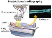
Much of the visualizing instruments that are used for medical diagnostics are derived or related to media technologies. The invention of the x-ray film in the 1950’s that was intended to record a persons lungs, was made possible because of the invention on image intensifier which soon after boosted the production of television sets. Since Wilhelm Rontgen discovery of the x-ray machine, it became standard in clinical settings as well as being standard for all to use as it being a good visualizing starting point for diagnosing. Beginning of the twentieth century x-rays started to appear in cinemas as well as campaign ads (Van dijick, pg. 12). One example was in a tuberculosis-prevention ad where trying to show what tuberculosis does to the body and this form of showing the public what it does and how looking at the x-ray they can reflect to themselves to prevent from getting tuberculosis (Lerner, pg. 383). Ads these gave rise to public interest by ether being appealing to the viewer’s eyes or that of getting the public interested in medical issues and for ones own health.
Some say that the visualizing trend in relation to visualization of medicine was due to mass media exploiting the power of interesting images, but in reality the opposite effect occurred where doctors and hospitals used it as an opportunity to add more to their public relations from these fascinating bodily images. Not to long after he innovation of the x-ray, many media and medical technologies came together to try and display the inside human body. Since the discovery of the x-ray and its film, it inspired to start using cameras as surgical intervention and even to the point of where its standard In most operating rooms. It also leads to the development of surgical instruments with cameras so they can visualize the inside of the body and what they are operating on (Van dijick, pg. 6).
Over the years the way the way we have looked at the body has changed. That when the first x-ray was invented it changed the entire perspective of how we look at human bodies and that it allows humans to see what is unseen.
This page has paths:
- Introduction Jamie DeBonis