Inside Decoys from Shelburne MuseumMain MenuDecoy MakingMakers represented in this projectDecoys included in this projectThe rationale behind which decoys were included in the project.About the RadiographsThe nitty gritty about how the radiography was done.BibliographyCited works and works that informed this project.About the AuthorsAbout Nancie Ravenel and Lesley Day MirlingNancie Ravenelbc84e2b969fab7c5f039797f42318c7fcfc8159bLesley Day Mirling3670b61b9eba655d6cf36db8e509081714fc05b1Shelburne Museum
A slice from a lateral view medical digital tomorgraph of a premier grade whistler drake decoy made by the Mason Decoy Factory, c. 1900
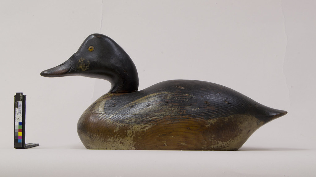
12017-08-04T13:08:08-07:00Nancie Ravenelbc84e2b969fab7c5f039797f42318c7fcfc8159b41362The image was taken at the Univeristy of Vemont Medical Center Hospital. Mason Decoy Factory (Detroit, Michigan, 1896-1924) Whistler Drake Decoy, ca. 1900 Wood, paint, and glass Museum purchase, acquired from Richard H. Moeller, 1956-707.118plain2017-08-05T13:53:28-07:001956-707.118Shelburne MuseumNancie Ravenelbc84e2b969fab7c5f039797f42318c7fcfc8159b
This page has annotations:
12017-08-04T13:21:54-07:00Nancie Ravenelbc84e2b969fab7c5f039797f42318c7fcfc8159bMark from outer cutting tooth on drill bitNancie Ravenel1plain2017-08-04T13:21:55-07:00Nancie Ravenelbc84e2b969fab7c5f039797f42318c7fcfc8159b
12017-08-04T13:22:39-07:00Nancie Ravenelbc84e2b969fab7c5f039797f42318c7fcfc8159bAnother mark from the center of a drill bit.Nancie Ravenel1plain2017-08-04T13:22:39-07:00Nancie Ravenelbc84e2b969fab7c5f039797f42318c7fcfc8159b
12017-08-04T13:21:18-07:00Nancie Ravenelbc84e2b969fab7c5f039797f42318c7fcfc8159bDrill bit centerNancie Ravenel1plain2017-08-04T13:21:18-07:00Nancie Ravenelbc84e2b969fab7c5f039797f42318c7fcfc8159b
12017-08-04T09:18:37-07:00Premier grade whistler drake decoy, 1956-707.1186Made c. 1900 by the Mason Decoy Factory, Detroit, MIplain2017-08-04T13:24:04-07:00 The radiographs show that the decoy is made of 3 pieces of wood, a head, an upper body and the lower body. The upper and lower body appear to have been carved as a single piece of wood and then split using a bandsaw, as suggested by the tool marks in the posterior-anterior image. The posterior-anterior image also shows that the glass eyes are enclosed within a metal jacket.
The lateral radiographic image shows that the body was hollowed using what appears to be a Forstener bit, and that the two halves of the body are secured with adhesive and finishing nails. The head is secured to the body with a wood dowel secured with a metal pin in the neck, as seen in the lateral image.
The lower half of the body also appears to have been hollowed using a Forstener bit applied with a wagging or angled motion to speed clearance, as evidenced by the shapes of the drill bit mark centers in a slice from a medical digital tomograph scan in the axial direction. Lateral slices from the digital tomograph scan provide an even clearer view of the bit mark, with a pointed center and a cutting tooth on the outer perimeter of the bit.
Digital medical tomograph take an x-ray image in a 10 degree sweep. The computer divides the resulting signal into slices. Features that are in the same plane as the slice are in focus. Those that are not appear blurry and less visible. This allows us to see beyond some of the features that are visible in the standard radiograph.