Scalar's 'additional metadata' features have been disabled on this install. Learn more.
Inside Decoys from Shelburne MuseumMain MenuDecoy MakingMakers represented in this projectDecoys included in this projectThe rationale behind which decoys were included in the project.BibliographyCited works and works that informed this project.About the AuthorsAbout Nancie Ravenel and Lesley Day MirlingNancie Ravenelbc84e2b969fab7c5f039797f42318c7fcfc8159bLesley Day Mirling3670b61b9eba655d6cf36db8e509081714fc05b1Shelburne Museum
About the Radiographs
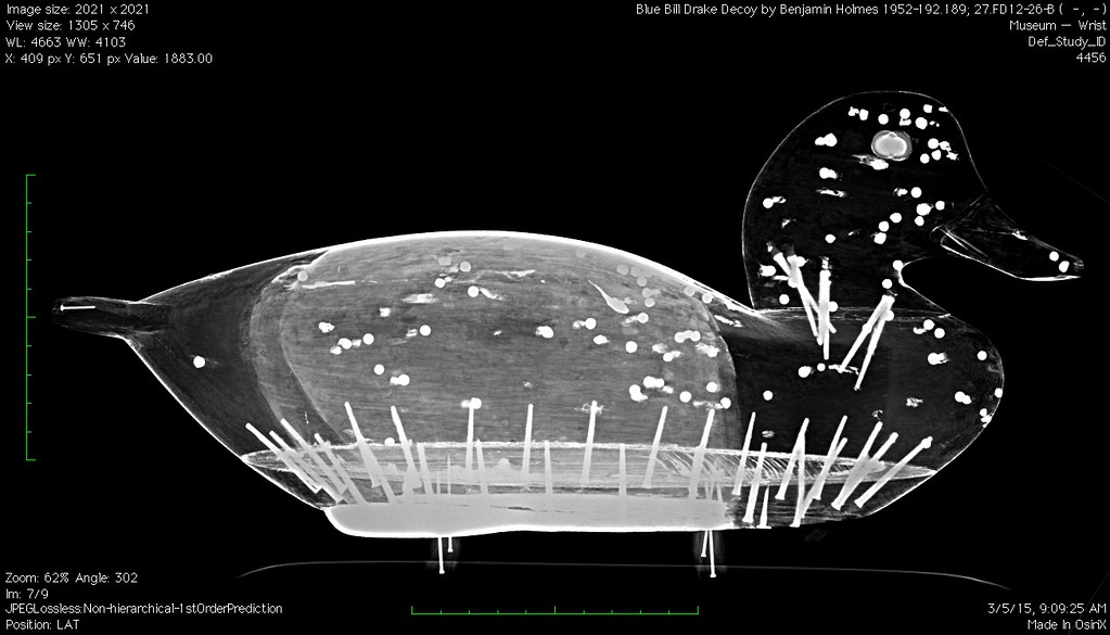
12015-02-01T12:19:32-08:00Nancie Ravenelbc84e2b969fab7c5f039797f42318c7fcfc8159b413611The nitty gritty about how the radiography was done.image_header2017-08-04T09:01:53-07:00Nancie Ravenelbc84e2b969fab7c5f039797f42318c7fcfc8159bThree different radiological techniques were used in the course of the project and were undertaken at the University of Vermont Medical Center's Radiology Department, facilitated by Michael Blakeslee. Standard radiography and Volume RAD tomosynthesis were undertaken using a GE Discovery XR656 machine by Carol Kittredge. Computed tomography (CT) was produced using a Phillips Ingenuity CT and the resulting files were processed by Heidi Streeter prior to them coming to the Shelburne Museum. Nancie Ravenel and Lesley Day Mirling viewed and further processed the resulting DICOM files using Adobe Photoshop, OsiriX (32 bit), or Horos to produce the jpeg images used for this site. Processing included re-sizing and re-orienting images and adjusting brightness to highlight features that would have been difficult to see otherwise. Videos and 2D orthogonal multi-planar reconstructions of the CT stacks were also produced using OsiriX Lite. Metadata regarding the specific objects imaged was also enhanced for image preservation purposes.
This page has paths:
12015-01-27T03:34:06-08:00Nancie Ravenelbc84e2b969fab7c5f039797f42318c7fcfc8159bInside Decoys from Shelburne MuseumNancie Ravenel52What's this all about?image_header1189402017-09-11T06:34:37-07:00Nancie Ravenelbc84e2b969fab7c5f039797f42318c7fcfc8159b